
Anatomy and physiology are fundamental sciences studying the structure and function of the human body, exploring cell, tissue, and system interactions, essential for understanding life and health․
1․1․ Definition and Importance of Anatomy and Physiology
Anatomy is the scientific study of the body’s structure, focusing on the organization and relationships of organs, tissues, and cells․ Physiology examines the functions and processes that enable the body to maintain life and health․ Together, they provide a comprehensive understanding of how the body works, from cellular mechanisms to system-level interactions․ These sciences are vital in medicine, healthcare, and biological research, offering insights into normal and pathological conditions․ Studying anatomy and physiology is essential for understanding human health, diagnosing diseases, and developing treatments, making them foundational for students, researchers, and professionals in the medical field․
1․2․ Interrelationship Between Anatomy and Physiology
Anatomy and physiology are deeply interconnected, as the structure of body parts determines their functions․ Anatomy provides the framework for understanding how tissues and organs are organized, while physiology explains how these structures work together to maintain life․ For example, the heart’s muscular walls (anatomy) pump blood (physiology) to meet the body’s needs․ This interrelationship is crucial for diagnosing and treating diseases, as changes in structure often lead to functional disorders․ Studying anatomy and physiology together offers a holistic understanding of the body, essential for advancing medical knowledge and improving healthcare practices․
1․3․ Branches of Anatomy and Physiology

Anatomy and physiology are divided into various branches, each focusing on specific aspects of the body․ Anatomy includes gross anatomy (study of visible structures), histology (microscopic study of tissues), and neuroanatomy (study of the nervous system)․ Physiology branches into areas like neurophysiology (function of the nervous system) and pathophysiology (study of diseases)․ Additionally, applied fields like comparative anatomy (study of different species) and vegetable anatomy (plant structure) exist․ These branches provide a comprehensive understanding of the body, aiding in medical advancements and practical applications in healthcare and research․

Histology: The Microscopic Study of Tissues
Histology involves the microscopic examination of tissues to understand their structure, function, and organization․ It is crucial for identifying tissue types and their roles in maintaining bodily functions․
2․1․ Types of Tissues in the Human Body
The human body is composed of four primary types of tissues: epithelial, connective, muscle, and nervous․ Epithelial tissues form linings and coverings, such as skin and mucous membranes․ Connective tissues provide support, structure, and connectivity, including bones, cartilage, and blood․ Muscle tissues are specialized for contraction, enabling movement and maintaining posture․ Nervous tissues facilitate communication through electrical and chemical signals, forming the basis of the nervous system․ Each tissue type has distinct functions and characteristics, working together to maintain overall bodily functions and health․ Understanding these tissues is foundational for studying anatomy and physiology, as they form the building blocks of organs and systems․
2․2․ Functions and Characteristics of Epithelial, Connective, Muscle, and Nervous Tissues
Epithelial tissues protect surfaces, line cavities, and regulate exchange of substances․ Connective tissues support, connect, and cushion organs, with examples like bones, cartilage, and blood․ Muscle tissues contract to enable movement, with types including skeletal, smooth, and cardiac․ Nervous tissues transmit and process information through electrical and chemical signals․ Each tissue has unique functions and structural adaptations, such as epithelial tight junctions or muscle fibers, essential for maintaining homeostasis and enabling bodily functions․ Understanding their roles and characteristics is crucial for comprehending human anatomy and physiology, as they form the foundation of organ systems and their interactions․

Levels of Structural Organization in the Human Body
The human body is organized hierarchically, starting from cells forming tissues, tissues forming organs, and organs functioning as systems, creating a complex, interdependent structure․
3․1․ From Cells to Organ Systems
The human body is organized in a hierarchical manner, beginning with cells as the basic structural and functional units․ Cells with similar functions group together to form tissues, such as epithelial, connective, muscle, and nervous tissues․ Tissues combine to create organs, which are specialized structures like the heart or liver․ Organs work together in coordinated systems, such as the digestive, respiratory, or circulatory systems, to perform complex functions essential for life․ This progression from cells to organ systems illustrates the intricate organization necessary for maintaining overall health and enabling the body to function as a unified whole․
3․2․ Understanding the Hierarchy of Body Structures
The human body is organized into a structural hierarchy that ensures efficiency and coordination․ At the most basic level, cells form tissues, which are specialized for specific functions․ Tissues combine to create organs, such as the heart or lungs, designed for precise roles․ Organs then function together within organ systems, like the circulatory or respiratory systems, to perform complex processes․ This hierarchical organization allows the body to operate as an integrated whole, with each level contributing to overall health and function․ Understanding this framework is essential for studying anatomy and physiology, as it explains how structures interact to maintain life and enable bodily functions․
Anatomical Terminology
Anatomical terminology includes directional terms like anterior, posterior, superior, and inferior, and body planes such as sagittal, frontal, and transverse, essential for precise communication and study․
4․1․ Directional Terms and Body Planes
Directional terms and body planes are essential for describing the location and orientation of structures within the body․ Terms like anterior (front), posterior (back), superior (above), and inferior (below) provide precise language for spatial relationships․ Body planes, such as the sagittal (divides the body into left and right), frontal (divides into front and back), and transverse (horizontal plane), help visualize the body in sections․ These terms and planes are critical for accurate communication in anatomy and physiology, enabling professionals to locate structures and describe movements effectively․ Mastery of this terminology is fundamental for understanding anatomical positions and relationships․
4․2․ Understanding Anatomical Positions
The anatomical position is a standardized reference point for describing body structures․ It involves standing upright with arms at the sides, palms facing forward, and feet parallel․ This position ensures consistency in describing movements and locations․ Key terms include flexion (bending), extension (straightening), abduction (movement away from the midline), and adduction (movement toward the midline)․ Understanding these positions and movements is vital for accurate communication in anatomy and physiology, aiding in the precise location of structures and the description of body dynamics․ This foundational knowledge is essential for healthcare professionals and students alike․

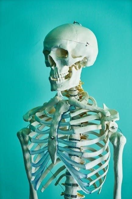
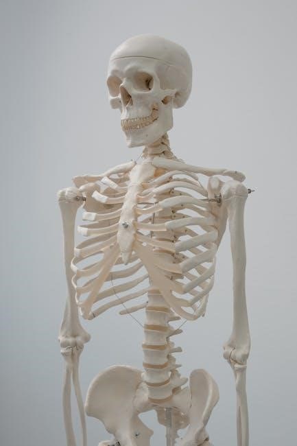
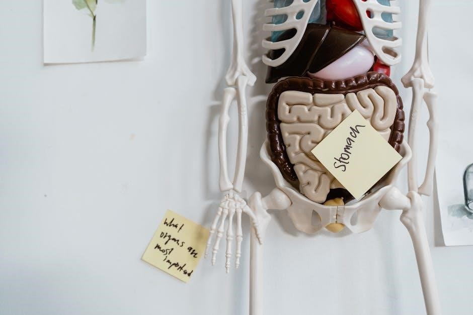
Skeletal System Overview
The skeletal system forms the body’s framework, protecting organs and facilitating movement․ It comprises 206 bones, divided into axial and appendicular skeletons, producing blood cells and storing minerals․
5․1․ Structure and Function of Bones
Bones are dynamic, living tissues that provide structural support, protect internal organs, and facilitate movement․ They consist of a dense outer layer (periosteum) and an inner spongy structure․ Compact bone offers strength and protection, while cancellous bone, with its trabeculae, reduces weight․ Bones also produce blood cells in marrow and store minerals like calcium․ The skeletal system’s integrity relies on bone tissue, composed of osteoblasts (bone-forming cells) and osteoclasts (bone-resorbing cells)․ This continuous remodeling process ensures bone health and calcium balance, essential for the body’s overall functionality and mobility․
5․2․ Classification of Joints and Their Importance
Joints, or articulations, are points where bones connect, enabling movement and stability․ They are classified into three main types: synarthroses (immovable), amphiarthroses (slightly movable), and diarthroses (freely movable)․ Synarthroses, like skull sutures, provide structural support․ Amphiarthroses, such as intervertebral discs, allow limited movement․ Diarthroses, including hinge and ball-and-socket joints, facilitate a wide range of motion․ Understanding joint classification is crucial for comprehending movement mechanics, treating injuries, and appreciating the skeletal system’s adaptability․ This knowledge aids in diagnosing joint-related disorders and enhancing mobility in clinical and therapeutic settings, making it a cornerstone of anatomy and physiology studies․
5․3․ Axial vs․ Appendicular Skeleton

The human skeleton is divided into the axial and appendicular systems․ The axial skeleton includes the skull, vertebral column, ribs, and sternum, forming the body’s central axis and protecting vital organs like the brain and heart․ The appendicular skeleton comprises the upper and lower limbs, shoulders, and pelvis, enabling movement and mobility․ The axial skeleton provides structural support and stability, while the appendicular skeleton facilitates locomotion and interaction with the environment․ Understanding their roles and functions is essential for analyzing posture, movement, and the overall integrity of the musculoskeletal system in anatomy and physiology studies․
Muscle Physiology
Muscle physiology examines the function and structure of skeletal, smooth, and cardiac muscles, enabling voluntary movement, support, and bodily functions through contraction mechanisms linked to the nervous system․
6․1․ Types of Muscles: Skeletal, Smooth, and Cardiac
There are three primary types of muscles in the human body: skeletal, smooth, and cardiac․ Skeletal muscles are voluntary, attached to bones, and enable movement․ Smooth muscles are involuntary, found in internal organs, and control processes like digestion․ Cardiac muscle is specialized for the heart, ensuring consistent blood circulation․ Each type has distinct structures and functions, with skeletal muscles controlled by the nervous system, smooth muscles operating autonomously, and cardiac muscles featuring intercalated discs for synchronized contractions․ Understanding their roles is essential for grasping movement, organ function, and cardiovascular health․
6․2․ Mechanism of Muscle Contraction
Muscle contraction occurs through the sliding filament theory, where actin and myosin filaments slide past each other․ It begins with a nerve impulse triggering the release of calcium ions from the sarcoplasmic reticulum․ These ions bind to troponin, shifting tropomyosin to expose myosin binding sites on actin․ The myosin heads then bind to actin, forming cross-bridges․ Using energy from ATP, the myosin heads pivot, pulling actin filaments toward the center of the sarcomere, resulting in contraction․ This process is essential for movement, maintaining posture, and regulating body functions․
Nervous System Basics
The nervous system controls voluntary actions, regulates involuntary functions, and enables communication through electrical and chemical signals․ It consists of the central and peripheral nervous systems, working harmoniously․
7․1․ Structure and Function of Neurons
Neurons, or nerve cells, are specialized cells designed to transmit information through electrical and chemical signals․ They consist of dendrites, a cell body, and an axon․ Dendrites receive signals from other neurons, while the cell body contains the nucleus and organelles essential for cell function․ The axon carries signals away from the cell body to synapses, where chemical neurotransmitters communicate with other neurons or target cells․ This structure allows neurons to process and transmit information, enabling functions like movement, sensation, and thought․ Understanding neuron structure and function is crucial for studying nervous system operations and disorders․
7․2․ Overview of the Central and Peripheral Nervous Systems
The central nervous system (CNS) includes the brain and spinal cord, functioning as the control center for the body by processing information and directing responses․ The peripheral nervous system (PNS) comprises nerves connecting the CNS to other body parts, enabling communication and sensory input․ The CNS interprets signals, while the PNS transmits and receives them․ Together, they coordinate voluntary and involuntary actions, ensuring the body operates efficiently․ Understanding their roles is essential for comprehending nervous system functions and diagnosing disorders․